Module 1: Introduction to Point-of-Care Ultrasound
From the Authors
The goal of this module is to cover some of the basics of ultrasound that are important to understand before moving on to the other modules. The introductory module will focus on high-yield topics such as basic concepts, terminology, artifacts, ultrasound probe motions, and scanning ergonomics.
—Ryan Paulus, DO, Nicole Yedlinsky, MD, CAQSM, FAAFP, RMSK, and Ben Clements, MD
Learning Objectives
After completing this module, the learner should be able to:
- Show a basic understanding of POCUS applications
- Select the correct probe for the examination
- Know what the different ultrasound modes are
- Describe important concepts on knobology
- Describe ultrasound imaging and probe motion terminology
- Use proper ultrasound ergonomics and positioning
- Identify common ultrasound artifacts
Module 2: Pulmonary Ultrasound
From the Author
Welcome to the Pulmonary Ultrasound module! My name is Ryan Paulus. I am an assistant professor and core faculty at the University of North Carolina Chapel Hill family medicine residency program. I integrate ultrasound into my daily practice and consider pulmonary ultrasound one of my favorite modalities. During this module, we will discuss performing the pulmonary exam and diagnosing pneumonia with ultrasound. —Ryan Paulus, DO
Learning Objectives
After completing this module, the learner should be able to:
- Perform an 8-point lung exam
- Identify findings of aerated lung ultrasound
- Interpret ultrasound findings consistent with pneumonia and pulmonary edema
- Incorporate lung ultrasound education in clinical precepting
Module 3: Soft-Tissue Ultrasound
From the Author
Welcome to the Skin and Soft Tissue Ultrasound module! My name is Nicoll Capizzano. I am a Clinical Assistant Professor and Clinical Ultrasound Director at Michigan Medicine, Family Medicine Department. During this module, we'll be looking at the case for using ultrasound prior to performing an I & D, ultrasound technique for soft tissue, demonstrate how to recognize normal versus abnormal tissue, and how to read the ultrasound scans. —Juana Nicoll Capizzano, MD
Learning Objectives
After completing this module, learners should be able to:
- Differentiate between the evidence behind most skin and soft tissue conditions for which point-of-care ultrasound can be used to help improve care
- Identify the general principles of image acquisition
- Recognize how to use point-of-care ultrasound in the evaluation of skin and soft tissue pathology
Module 4: Renal/Bladder Ultrasound
From the Author
Welcome to the Renal and Bladder Ultrasound module! My name is Ben Clements and I am an assistant professor at the University of Vermont Larner College of Medicine. I am the director of our Point of Care Ultrasound Program for the Department of Family Medicine and the POCUS Curricular Lead for the UVM Family Medicine Residency. In this module, we will discuss performing the renal and bladder exam, focusing on identifying hydronephrosis. The module incorporates a case of a resident using point-of-care ultrasound to diagnose urolithiasis. —Ben Clements, MD
Learning Objectives
After completing this module, the learner should be able to:
- Perform bilateral, short and long access renal exam
- Perform bladder exam
- Interpret ultrasound findings consistent with hydronephrosis
- Differentiate severity of hydronephrosis
- Understand when renal ultrasound prompts further evaluation
- Incorporate renal ultrasound education in clinical precepting
Module 5: Deep Venous Thrombosis (DVT)
From the Author
Welcome to the Performing the DVT Exam module. My name is Noah Furr, MD, out of Nellis Air Force Base. I'm a family physician on the faculty at the residency program there. The purpose of this module is to teach you how to do the Deep Vein Thrombosis (DVT) exam. You won't see all the bad stuff you might find, but you will learn where you need to go and what you need to see in order to call the exam. —Noah Furr, MD
Learning Objectives
After completing this module, the learner should be able to:
- Perform a 2-region compression exam to evaluate for deep venous thrombosis with POCUS
- Know indications for DVT evaluation with POCUS and differences/indications for variations of DVT US and adjunctive lower extremity imaging
- Understand normal findings of the lower extremity deep venous system
- Interpret ultrasound findings of a deep venous thrombosis
- Identify clinical scenarios, limitations, and pitfalls for DVT POCUS exams
Module 6: Musculoskeletal Ultrasound
From the Author
In this module, we want you to come away knowing the basics of performing Musculoskeletal (MSK) ultrasound, with an emphasis on being able to identify the appearance of normal MSK targets. MSK ultrasound is useful to identify pathology such as fractures, joint effusions, nerve entrapments, and tendon damage. We can also use MSK ultrasound for procedural guidance, such as joint aspirations and injections. —Nicole Yedlinsky, MD, CAQSM, FAAFP, RMSK
Learning Objectives
After completing this module, the learner should be able to:
- Identify sonographic appearance of MSK targets
- Discuss limitations of MSK ultrasound
- Use POCUS to identify MSK pathology
- Demonstrate principles of US guidance for anesthesia, injections, and aspiration
Module 7: Gallbladder Ultrasound
From the Author
Welcome to the Gallbladder module! My name is Puja Dalal. I am an assistant professor and the Director of POCUS at the Novant Health Family Medicine Residency Program in Cornelius, North Carolina. The purpose of this module is to teach you how to obtain images of the gallbladder. It covers various approaches for locating the gallbladder and provides tips for obtaining optimal images. The module concludes with a case of a resident using POCUS to diagnose cholelithiasis. - Puja Dalal, MD, FAAFP
Learning Objectives
After completing this module, the learner should be able to:
- Perform a limited gallbladder exam
- Identify normal gallbladder on ultrasound
- Interpret ultrasound findings consistent with cholelithiasis
- Understand ultrasound findings consistent with cholecystitis
- Incorporate gallbladder ultrasound education in clinical precepting
Module 8: OB/Gyn Ultrasound
From the Author
The goals of this module are to develop the essential skills to ask the basic questions – is there an intrauterine pregnancy and, if so, is it viable. Similarly, if there is no intrauterine pregnancy are there any overt signs of ectopic pregnancy or rupture that require immediately management or simply close follow up. As always, it is recommended that, after the basics are obtained, the provider dedicate some time reading about a detailed first trimester exam, if only to understand the possibilities and limits of POCUS. —Brandon Williamson, MD, FAAFP, RDMS
Learning Objectives
After completing this module, the learner should be able to:
- Perform a transabdominal exam of a first trimester pelvis
- Identify findings of a viable intrauterine pregnancy (IUP)
- Interpret ultrasound findings consistent with an extrauterine pregnancy
- Interpret ultrasound findings of a missed abortion
- Incorporate first trimester ultrasound in clinical precepting
Module 9: Abdominal Aortic Aneurysm (AAA) Screening
From the Author
Welcome to the AAA module. My name is John Doughton. I am an assistant professor at the University of North Carolina Chapel Hill family medicine residency program and director of the POCUS curriculum for the UNC School of Medicine. I also work with our rural residency track, and my outpatient work occurs in the FQHC setting. My patients experience many barriers to care, and it can be very difficult to get them screening exams. I believe that bedside ultrasound can expand access and improve care. During this module, we will discuss how to teach and perform a screening AAA exam. —John Doughton, MD
Learning Objectives
After completing this module, the learner should be able to:
- Perform a screening AAA exam
- Highlight areas of highest importance for detecting a AAA
- Obtain measurements at the proximal, mid, and distal aorta
- Recognize fusiform and saccular aortic aneurysms
- Identify common pitfalls to visualizing the abdominal aorta
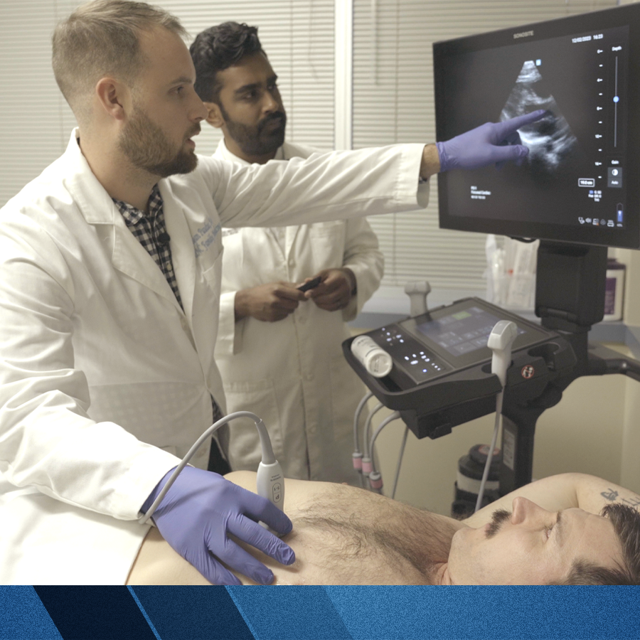
Module 10: Cardiac Ultrasound
From the Author
Performing a cardiac point-of-care ultrasound requires both didactic education and hands-on learning. I often find it most successful if learners have a quick lecture, prior to a workshop, or scanning to understand the anatomy and spatial positioning of structures. The probe positions in cardiac ultrasound are quite different, and therefore it can be useful to review probe orientations prior to walking into a patient’s room and scanning. —Hiten Patel, MD
Learning Objectives
After completing this module, the learner should be able to:
- Obtain four cardiac windows (parasternal long, parasternal short, apical 4 chamber, and subxiphoid)
- Know the indications of cardiac ultrasound
- Understand the normal findings of a heart ultrasound
- Interpret ultrasound findings seen in heart failure
- Identify clinical scenarios in which POCUS can help with clinical care